Day 1 :
Keynote Forum
James L. Sherley
Asymmetrex, LLC,
USA
Keynote: Current driving factors in stem cell-based regenerative medicine
Time : 09:00-09:25
Biography:
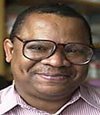
James Sherley graduated from Harvard College (1980) and completed joint M.D./Ph.D. degrees at the Johns Hopkins University School of Medicine (1988). After post-doctoral studies at Princeton University, beginning in 1991 he led cancer cell molecular biology research at Fox Chase Cancer Center. In 1998, he began adult stem cell research at Massachusetts Institute of Technology, and in 2007 continued at Boston Biomedical Research Institute. In 2013, he founded the Adult Stem Cell Technology Center, LLC, (ASCTC), which he currently directs. ASCTC is developing technologies for mass-producing human tissue stem cells for applications in drug development and transplantation medicine.
Abstract:
Modern medicine is now experiencing a deluge of unproven regenerative medicine treatments. Although there is considerable concern among stem cell scientists and physicians that many new cell therapies occur without the approval and supervision of government agencies like the Food and Drug Administration in the U.S., many new treatments are, in fact, being evaluated in certified clinical trials. However, even in the case of certified trials, there is ample reason to be circumscribed in expectations for them to yield meaningful progress. Because of patients’ and patient advocate groups’ high level of hope for relief from otherwise intractable and incurable ailments and disorders, the field of regenerative medicine is now propelled to act expediently to deliver the promises of stem cell medicine. This volatile situation engenders a high risk for the development of scientifically pre-mature clinical trials. In particular, cell transplantation trials driven more by factors of technical feasibility than by factors of biological plausibility should provoke concern regarding their likelihood to contribute to the success of regenerative medicine.
Keynote Forum
Ornella Parolini
President
IPLASS(International Placenta Stem Cell Society)
Italy
Keynote: In regenerative medicine does stem cell differentiation really matter?
Time : 09:25-09:50
Biography:
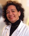
Ornella Parolini is the Director of E. Menni Research Center, Fondazione Poliambulanza, Brescia, Italy and the President of IPLASS - International Placenta Stem Cell Society. She has pioneered research on human placenta-derived stem cells. Her research is focused on the immunomodulatory, anti-inflammatory and anti-fibrotic properties of amniotic and chorionic membrane-derivatives as amnion patches, amniotic cell subpopulations and their conditioned medium, as products applicable in regenerative medicine. She is author of over 100 publications in peer-reviewed journals as well as several book chapters. She also has several patent applications
Abstract:
Stem cells act as progenitors for multiple other cell types and also contribute to tissue homeostasis, thus suggesting that these cells could be exploited for tissue regeneration and loss of organ function. This concept began to take shape with the discovery of cells such as embryonic stem cells and hematopoietic stem cells, which embody the classical features of stem cells, i.e. the ability to proliferate almost indefinitely and differentiate into multiple lineages. Combined with the ability to engraft, these features open the possibility to reconstitute tissues/organs by generating tissue-specific mature cells through differentiation. Mesenchymal stem/stromal cells (MSCs) very likely possess the same characteristics as other stem cells, although with fundamental aspects of their biology yet to be determined due to the confounding aspect of their intrinsic heterogeneity as a cell population. Recent evidence suggests that the factors released from these cells, rather than their intrinsic differentiation capacity, are the key players underlying their actions in vivo, ultimately raising questions relating to the use of effector molecules for therapeutic purposes. Our studies have mostly been focused on mesenchymal stem/stromal cells isolated from a human term placenta tissues, such as the amniotic membrane (hAMSC). We have demonstrated a paracrine action of hAMSC by showing a significant disease attenuation through the application of cells or their conditioned medium, and even amniotic membrane patches, in animal models of several inflammatory-associated diseases, such as in animals with ischemic hearts, lung and liver fibrosis, and rheumatoid arthritis. Altogether, these studies highlight the potential use of hAMSC and their derivatives (e.g. amniotic patches and conditioned medium), as a therapeutic tool in degenerative diseases based on altered immune reactions and inflammatory processes. In this presentation we will discuss evidence suggesting that a major role for paracrine actions of these cells, rather than their differentiation, underlies their therapeutic effects.
- Workshop on: Athletes, OA, and Regenerative Medicine Â
Location: Vienna Room
Session Introduction
Susan Chubinskaya
Rush University Medical Center
USA
Title: Post-traumatic osteoarthritis: clinical reality and experimental approaches
Time : 09:50-10:15
Biography:
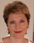
Susan Chubinskaya, PhD, The Ciba-Geigy Professor of Biochemistry; has a primary appointment in the Department of Biochemistry with conjoint appointments as Professor of Internal Medicine (Section of Rheumatology) and Orthopedic Surgery at Rush University Medical Center. She is also Associate Provost for Academic Affairs at Rush. Her research interests are the growth factors and the mechanisms of cartilage degeneration and repair in aging and post-traumatic osteoarthritis. As a Principal Investigator, she secured more than $4.5M (direct cost) of extramural funding, presented more than 140 invited and podium lectures, published 80 peer-reviewed manuscripts, 10 book chapters, and more than 180 peer-reviewed abstracts.
Abstract:
Post-traumatic or early osteoarthritis (PTOA) occurs as a result of joint overuse or joint injury and primarily affects young people, actively involved in various sports. They are eager to return to regular physical routines as soon as possible, which may lead to re-injury or more severe traumas. In acute joint injuries, cartilage damage likely occurs at the time of the initial trauma or soon after. Therefore, understanding how to prevent cartilage damage before irreversible progression begins, may provide a window of opportunity for the evaluation of risk and diagnosis of early PTOA, which takes years to develop. By the time it becomes symptomatic, only surgical interventions remain as a viable treatment option. There is a critical need to develop strategies for improving the outcomes after surgical interventions that would delay or prevent the subsequent development of PTOA. Treatment of the pre-osteoarthritic joint disease is a new concept, which emphasizes the need for preventive strategies that will modify the course of a disease. The current approach to the clinical treatment of OA is the palliation of symptoms arising from late-stage disease. Early-stage disease or pre-osteoarthritis disease is clinically silent in that structural changes typically precede clinical signs and symptoms of pain, deformity, functional limitations, and disability. Metabolic changes in articular cartilage, synovium, and subchondral bone may represent the earliest measurable changes in pre-OA conditions. As such, it may lead to new intervention strategies that support the development of disease-modifying therapies. In this presentation we will review the latest advances in biologic approaches to cartilage repair.
Dennis M Lox
Sports and Regenerative Medicine Centers
USA
Title: Athletes, sports medicine and stem cells: Regenerative applications
Time : 10:15-10:35

Biography:
Dennis M Lox, MD is the Director of the Sports and Regenerative Medicine Centers A Stem Cell Center of Excellence in the Washington D.C. Metro Area and the Tampa Bay Metro Area. He attended the University of Arizona, where he was Phi Beta Kappa studying Liberal Arts. He then completed his medical training at Texas Tech University, followed by his residency program in PM&R at The University of Texas Health Science Centers at San Antonio. He has always pursued Sports Medicine, and this led to a path into Regenerative Medicine. He has edited 2 medical texts, 9 book chapters, and lectures national and internationally on sports, regenerative medicine and stem cells.
Abstract:
Athletic endeavor has always intertwined with pursuing optimal physical performance. The inherent risk of traumatic injury has placed sports medicine at the forefront of progressive treatment. The emergence of Regenerative Medicine has led to the clinical translation of sports medicine related problems. The use of Biologics, Platelet Rich Plasma (PRP) and Stem Therapy are foundations for this approach. The scientific literature will be explored to provide a foundation for the pathophysiology of injury and trauma as it relates to sports medicine, and the regulation of catabolic responses through cellular signaling and cytokines. The rationale for Regenerative Medicine applications such as stem cell treatments in sports medicine is presented. Various clinical cases are presented to illustrate the utilization of stem cell therapy and PRP as a therapeutic strategy to assist athletes in their return to sport. Select situations in which standard medical treatment is a surgical intervention, may be unsuitable for return to sport. A Regenerative Medicine treatment model incorporating Stem Cell Therapy, may provide suitable alternative strategies that facilitate healing and repair, without precluding return to sport. In the sports medicine world success is measured by return to sport. The clinical translation of Stem Cell Therapy into sports medicine may foster a transformation of treatment strategy.
- Symposium On Stem Cell Medical Engineering
Location: Vienna Room
Session Introduction
James L. Sherley
Founder and Director
Asymmetrex, LLC
Title: Asymmetric Self-Renewal: The fundamental property of tissue stem cells is the fundamental principle for their medical engineering
Time : 10:35-10:55
Biography:
James Sherley graduated from Harvard College (1980) and completed joint M.D./Ph.D. degrees at the Johns Hopkins University School of Medicine (1988). After post-doctoral studies at Princeton University, beginning in 1991 he led cancer cell molecular biology research at Fox Chase Cancer Center. In 1998, he began adult stem cell research at Massachusetts Institute of Technology, and in 2007 continued at Boston Biomedical Research Institute. In 2013, he founded the Adult Stem Cell Technology Center, LLC, (ASCTC), which he currently directs. ASCTC is developing technologies for mass-producing human tissue stem cells for applications in drug development and transplantation medicine.
Abstract:
Tissue stem cells do more than “self-renew and differentiate.†In fact, this commonly applied description may often be erroneous. Some tissue stem cells may never differentiate. Instead they divide to produce lineage-committed differentiating cells while retaining their own stem cell state. This remarkable capability is asymmetric self-renewal. Asymmetric self-renewal may be accomplished by either of two distinctive types of cell kinetics programs. Individual tissue stem cells may undergo determined asymmetric cell divisions that produce a stem cell sister and a lineage-committed sister; or small pools of stem cells may divide to produce stochastically either two sister stem cells or two lineage-committed cells. Both cell kinetics programs can achieve the same essential feature of vertebrate tissues, mature cell differentiation without loss of the cell development program. Increased application of the principle of tissue stem cell asymmetric self-renewal will accelerate success in engineering of human tissue stem cells for medical innovations. Asymmetric self-renewal poses an intrinsic challenge to production of tissue stem cells for use in cell replacement therapies like diabetes, anemias, liver failure, and tissue injuries. As a unique cellular process that defines tissue stem cells universally, asymmetric self-renewal is an ideal biomarker for quantitative dosing of tissue stem cells. Similarly, the unique cell kinetics output of tissue stem cells projects a unique cell growth signature that can be used to monitor tissue stem cells for novel drug evaluation assays. Such recent advances in tissue stem cell medical engineering based on the fundamental principle of asymmetric self-renewal will be presented.
Coffee Break 10:55-11:10 @ Vienna Foyer
- Track 3: Stem-Cell Transplantation
Track 7: Cell Mechanics
Track 8: Cell Based Therapies of Blood Diseases
Location: Vienna Room
Chair
James L. Sherley
Asymmetrex LLC, USA
Co-Chair
Ornella Parolini
International Placenta Stem Cell Society (IPLASS), Italy
Session Introduction
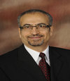
Shirwan Haval
Professor
University of Louisville
USA
Title: ProtExâ„¢ technology as an effective means of engineering bone marrow cells with immunoregulatory molecules for the establishment of mixed hematopoietic chimerism
Time : 11:10-11:30
Biography:
Dr. Shirwan is Dr. Michael and Joan Hamilton Endowed Chair in Autoimmune Disease, Professor of Microbiology and Immunology, Director of Molecular Immunomodulation Program at the Institute for Cellular Therapeutics. He conducted his graduate studies at the University of California in Santa Barbara, CA, and postdoctoral studies at California Institute of Technology in Pasadena, CA. He joined the University of Louisville in 1998 after holding academic appointments at various academic institutions in the United States. Dr. Shirwan’s research focuses on the modulation of immune system for the treatment of immune-based diseases with particular focus on type 1 diabetes, transplantation, and development of prophylactic and therapeutic vaccines against cancer and infectious diseases. Dr. Shirwan is an inventor on 16 worldwide patens, widely published, organized and lectured at numerous national/international conferences, served on study sections for various federal and non-profit funding agencies, and is on the Editorial Board of a dozen of scientific journals. He is member of several national and international societies and recipient of various awards.
Abstract:
Allogeneic bone marrow transplantation (BMT) has the potential to cure a series of inherited and acquired hematological disorders. The routine application of allogeneic BMT as a therapeutic intervention in the clinic, however, is complicated by nonmyeloablative conditioning of the recipient, graft-versus-host disease (GVHD), and lack of engraftment. Although elimination of T cells from the donor BM inoculum is effective in curtailing GVHD, it results in compromised engraftment. Thus, strategies targeting specific and effective elimination of only the pathogenic T cells may have important implications for routine application of BMT to the clinic. We have developed a novel approach, designated as ProtEx™, to engineer bone marrow cells with an exogenous protein immunoregulatory protein, SA-FasL, and tested the efficacy of the engineered cells to engraft in allogeneic recipients. Our results demonstrate the robust efficacy of this approach to establish mixed hematopoietic chimerism under nonmyeloablative conditioning and without complication of GVHD.
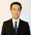
Gary Guishan Xiao
Director
Genomics and Functional Proteomics Laboratories
Creighton University
USA
Title: Regulation of miR-34a-SIRT1 axis reduced breast cancer stemness
Time : 11:30-11:50
Biography:
Dr. Xiao is a Professor and the Director in School of Pharmaceutical Science at Dalian University of Technology China and the Functional Genomics and Proteomics Laboratories at the Creighton University School of Medicine, and an internationally recognized expert in the field of genomics and proteomics of cancer and bone disease. Dr. Xiao earns his Ph.D. in molecular biology in Institute of Biophysics at Chinese Academy of Sciences. He had his postdoctoral research trained in Baylor College of Medicine and UCLA, focusing on pharmacokinetics and biochemistry of non-steroid inflammatory drugs, and cell cycle regulation. He has been regular reviewer or ad hoc reviewer for several medical journals, different funding agency and several journal Editorial Board members and senior Editors.
Abstract:
Introduction Recent studies showed that enforced expression of miR-34a resulted in elimination of cancer stem cells in many tumors, including prostate, pancreatic cancers. In this study, we aim to understand regulatory mechanism of miR-34a in breast cancer stem cells (BCSCs). Methods CD44+/CD24− breast cancer stem cells (BCSCs) were isolated from MCF-7 cell line. Total RNA and protein was extracted from BCSCs and xenograft tissues. Levels of miR-34a, Sirtuin1 (SIRT1), ALDH1, BMI1 and Nanog, were measured by using qRT-PCR and Western blot. Cell proliferation and differentiation were measured by assays of CCK-8, colony formation and mammosphere formation. Cell apoptosis was measured by flow cytometry. Four groups of mice (n=5), lentivirus-hsa-miR-34a, shRNA-SIRT1, lentivirus-hsa-NC, and shRNA-Control, were used for study on tumor formation and growth. Results Lower endogenous level of miR-34a and higher level of Sirtuin1 (SIRT1) gene in CD44+/CD24- BCSCs than in breast cancer cells was identified. Further study demonstrated that miR-34a directly targeted SIRT1 gene leading to reduced SIRT1 expression in the BCSCs. Either ectopic expression of miR-34a or silenced SIRT1 in MCF-7 cells inhibited cellular proliferation, and led to cell apoptosis. Overexpression of miR-34a also suppressed expression of ALDH1, BMI1 and Nanog, and decreased capacity of mammosphere formation of the BCSCs significantly, suggesting the reduced stemness of BCSCs. Studies in vivo showed that stable expression of miR-34a reduced tumor burden significantly in nude mice xenografts. Conclusion Our results sshowed that anti-synergetic regulation of miR-34a-SIRT1 axis affected proliferation, colony formation, and mammosphere formation in BCSCs, suggesting that miR-34a-SIRT1 axis may play an important role in self-renewal and stemness maintenance of BCSCs. This study may provide a novel BCSCs specific therapeutic strategy to improve breast cancer treatments.
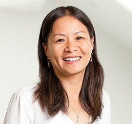
Xianmin Zeng
XCell Science Inc
California
Title: Pluripotent stem cell-based therapy for Parkinson’s disease
Time : 11:50-12:10
Biography:
Xianmin Zeng is a leading stem cell biologist with expertise in neural development of human ESC/iPSC. One of her research focuses is to study neural/neuronal development in human and to model neurodegenerative diseases, with a focus on Parkinson’s disease, using patient-specific and engineered isogenic iPSC lines. She has developed scalable processes of generating functional CNS cells and PNS cells for cell replacement therapy and screening drugs of neurotoxic and/or neuroprotective effects. She joined the faculty of the Buck Institute in 2005 where she builds the Institute’s Stem Cell Program. She received early tenure in 2009 and has been the Director of North Bay Shared Research Laboratory for Stem Cell and Aging at the Institute since 2008.
Abstract:
For cell-based therapy to treat Parkinson’s disease (PD), we have developed a Good Manufacturing Practice (GMP)-compatible process for generating authentic midbrain (A9/A10) dopaminergic neurons using defined media from either human embryonic stem cells (ESC) and induced pluripotent stem cells (iPSC). We have identified the optimal time point at which dopaminergic neurons can be frozen, shipped and thawed without compromising their viability and ability to mature in vivo after transplantation. We have also performed IND-enabling preclinical efficacy and safety studies using cells manufactured by this process. Our results show robust long-term survival of transplanted dopaminergic neurons and functional recovery at 6 months post transplantation in the 6-hydroxydopamine (6-OHDA)-lesioned rat PD model. To optimize the transplantation process we have helped develop and test a surgical delivery catheter that can be integrated with an US Food and Drug Administration (FDA)–approved iMRI skull-mounted aiming device and targeting software. We have demonstrated biocompatibility of the cells with the device and successfully delivered with iMRI guidance into swine models. In summary, 1) we have access to a clinical-grade ESC and iPSC line as well as their matched research grade differentiated cells; 2) We have identified a manufacturing site and have access to the DMF; 3) We have performed preclinical studies with cells manufactured by the GMP-compatible process we have developed and 4) We have coordinated with clinicians on developing a delivery procedure and a surgical trial design.
Margarita Glazova
Russian Academy of Sciences
Russia
Title: Intrinsic role of p53 in the differentiation of neural stem cells
Time : 12:10-12:30
Biography:
Margarita Glazova received her PhD (1997) from Sechenov Institute of Evolutionary Physiology and Biochemistry Russian Academy of Science. She performed Postdoctoral studies at Turku Centre for Biotechnology, University of Turku/ÅboAkademi, Finland (2001-2003) and then at East Carolina University Brody School of Medicine, Department of Physiology, Greenville NC, USA (2004-2006). Now, she is Senior Researcher and group leader at the Sechenov Institute of Evolutionary Physiology and Biochemistry, Russian Academy of Sciences, St.Petersburg, Russia. Her main interests concern the study of adult neurogenesis and neural stem cells differentiation at normal and under neuropathology disease, such as epilepsy. She has published more than 30 articles in peer-reviewed scientific journals.
Abstract:
p53 is a tumor suppressor protein that induces cell cycle arrest, senescence, or apoptosis in response to cellular stresses. Recent studies suggest the involvement of p53 also in the regulation of neural stem cell self-renewal and differentiation. However, our knowledge about molecular mechanisms of the p53-mediated neuronal differentiation is still limited. The purpose of this study was to reveal the signaling pathways involved in p53-mediated neuronal differentiation. Recently, we demonstrated that p53 deficiencies affected not only on the functional activity of mature neurons of hypothalamus, but also differentially changed the cell’s populations. Moreover, our and others data demonstrated that p53 can induce MAPK activity, the well-known pathway involved in NSC differentiation. Our study was performed on neural progenitors isolated from the hippocampus of new-born mice.The obtained data demonstrated that p53 acts on neural differentiation in cooperation with MAPK. Moreover, the results revealed that GSK3b also mediates the effects of p53. This work was supported by grant RFBR 13-04-01431-a.
Caroline Hoemann
Professor
Ecole Polytechnique
Canada
Title: Fusion peptide P15-CSP coatings suppress biofilm formation and advance mesenchymal stem cell osteogenic differentiation on hydrophilic but not hydrophobic surfaces
Time : 12:30-12:50
Biography:
Dr. Hoemann (PhD, MIT, 1992) is a full professor of Chemical Engineering and Biomedical Engineering at the Ecole Polytechnique in Montreal, Canada, and an FRSQ National Research fellow. She has published over 50 articles, 3 opinion papers and 6 patents in the area of tissue engineering, cartilage/bone repair, and blood/innate immune responses to biomaterials. She is a member of the Orthopedic Research Society, Fellow Member of the International Cartilage Repair Society, lead author on the ICRS recommendation paper, “International Cartilage Repair Society (ICRS) recommended guidelines for histological endpoints for cartilage repair studies in animal models and clinical trials, Cartilage 2011, 2:153-172” and serves on the editorial board of Cartilage, and The Open Orthopaedics Journal. She spent 5 years as Director of Cartilage Repair, in a Montreal-based biomedical device company, where she co-invented and co-developed a novel medical device for articular cartilage repair, BST-CarGel® that was tested in an 80-patient randomized controlled clinical trial, received CE-mark approval, and is now being used in European clinics. Her current research program has created novel bioengineered biomaterial blood clot implants, developed new methods for staining and analyzing human cartilage repair biopsies, and has made advances in understanding the role of therapeutic inflammation and subchondral bone remodeling in cartilage repair responses. Her translational research program aims to understand the influence of local inflammation on cartilage and bone repair, in order to bring new treatment options to patients with arthritis.
Abstract:
In periodontal bone reconstruction procedures, bone growth is enhanced by osteogenic factors and impaired by wound infection. PepGen P-15 (P15) is an osteogenic collagen-mimetic peptide, and competence stimulating peptide (CSP) is a cationic amphiphilic peptide with antimicrobial activity. This study analyzed whether surfaces coated with P15-CSP fusion peptide have dual anti-infective and pro-osteogenic activities in vitro. Plastic surfaces were dry-coated with or without P15-CSP, CSP, P15, or left uncoated, then inoculated with Streptococcus mutans or seeded with human mesenchymal stem cells (MSCs). CSP coatings inhibited S. mutans planktonic growth while P15-CSP coatings suppressed biofilm formation. P15 had no antimicrobial or anti-biofilm activity. MSCs adhered and spread more rapidly on P15-CSP and CSP coatings compared to P15-coated and uncoated culture wells, although CSP-coatings showed slight cytotoxicity to MSCs after 24 hours of culture. After 3 weeks in osteogenic culture, MSCs up-regulated alkaline phosphatase activity on tissue culture plastic and P15, CSP and P15-CSP coatings, with a selectively higher matrix mineralization on P15-CSP-coated surfaces (p<0.05). In other assays, peptides were adsorbed to surfaces pre-coated with thin films of plasma-polymerized hydrophilic carboxylated ethylene (L-PPE:O) or hydrophobic hexamethyl disiloxane HMDSO. MSCs expressed alkaline phosphatase activity on carboxylated surfaces with higher matrix calcificationwhen L-PPE:O was further coated with P15 or P15-CSP. Unexpectedly, MSCs cultured on hydrophobic thin films developed alkaline phosphatase activity and failed to calcify, with or without peptide coatings. This study revealed that P15-CSP coatings stabilized by electrostatic interactions exert dual properties of anti-biofilm activity, and accelerated MSC adhesion and end-stage osteogenic differentiation.
Francisco Ruiz-Navarro
Regmed Research Center
Austria
Title: Neuron point-of-care stem cell therapy (N-POCST): Transplantation of autologous bone marrow-derived stem cells for traumatic central nervous system disorders, the future of a new method
Time : 12:50-13:10

Biography:
Dr. Francisco Ruiz-Navarro (M.D) is Research Associate at Regmed Research Center in Vienna, Austria. Before, he was working as researcher at the Mexican Institute for Neurology and Neurosurgery in the Cerebrovascular Department focused in multi-centric population studies with Hispanic stroke patients. He was research assistant in the Center for Research and Advanced Studies of the National Polytechnic institute (CINVESTAV) in Mexico City at the Brain Bank and Physiology, Biology and Neuroscience Department. He was Research Assistant in La Raza Medical Center in Mexico City in the pediatric nephrology department. He obtained the Medical Degree in Anahuac University in Mexico City and became USMLE board certified in United States of America with outstanding grades. During his carrier he had been attending physician in different Mexican hospitals in Mexico City as part of general medicine, anesthesiology and neurosurgical teams. He performed clinical rotations at Jackson´s memorial Hospital Miami, USA in the stroke unit, neuroradiology department and neurosurgery department. His current research is directed to the use of autologous bone marrow-derived stem cells for neurological diseases.
Abstract:
Traumatic insults of the central nervous system (CNS) result in definite chronic disability that respond partially and only in certain cases to rehabilitation. The use of autologous bone marrow-derived stem cells (BMSC) has been suggested to be a feasible treatment that improves the neurological recovery in these disorders. In the acute phase the stem cells transplanted target is to prevent neurons death by different secreting factors that provide neuroprotection via improvement of the local microenvironment. In a chronic scenario the objective is the restoration of neural tissue by transdifferentiation. Although the acute and chronic treatment seemed to have positive results in preclinical and ongoing pilot studies, the rationale of neuroprotection makes more probably to achieve better outcomes with an acute intervention. In order to accomplish the intervention immediately after the neurologic insult a method to process bone BMSC in an effective and prompt way should be available. The Neuron point-of-care method (N-POCST) described before by our group for other neurological diseases like amyotrophic lateral sclerosis, is a method of on-site cell separation with a closed system that uses density centrifugation technique with a fixed 10% reduction rate to isolate bone marrow-derived mononuclear cells (BM-MNCs) immediately after bone marrow aspiration and without mayor cell modification. The BM-MNCs concentrate is infused intrathecal (IT) after the separation. Several publications had described the safety of intrathecal stem cells infusion, others described the positive results with autologous BMSC, other studies confirm the effectiveness of the cell separation process to concentrate BM-MNCs, all of these results together open the track to develop further randomized controlled clinical trials with N-POCST in patients with acute traumatic brain injuries.
Bruno Doiron
University of Texas Health Science Center at San Antonio
USA
Title: Uncomplicated way to induce pancreatic beta cell formation in vivo through “Cellular Networking, Integration and Processing (CNIP)
Time : 13:55-14:15
Biography:
Bruno Doiron, Ph.D is an Assistant Professor at the University of Texas Health Science Center at San Antonio. He received his undergraduate degree from University of Moncton, Canada and graduate degrees from University of Montreal, Canada and University of Paris Descartes, Paris, France. As project leader he has made major discoveries in the field of gene regulation by nutrients and has 4 patents on the modulation of glucose metabolism as it relates to the treatment diabetes and cancer. Dr. Doiron has extensive experience in basic research at the physiologic and molecular levels and in respective applications to the biotechnology field.
Abstract:
We create a novel approach in biology that integrates multiples levels of cellular physiology to regulate organ function. We demonstrated by using CNIP, we can increase the pancreatic beta cell number in vivo in adult animals. The CNIP approach dictates simultaneous intervention at three major levels in cell processing: first, at the level of gene expression; second, at the level of the membrane receptor; and third, at the level of intracellular metabolism. By targeting all three levels simultaneously, one can induce a synergistic interaction to produce the desired cellular effect, i.e. pancreatic beta cell formation. The CNIP approach would have an enormous impact on the treatment of type 1 and 2 diabetes, where one of the major physiopathologic abnormalities is the loss of pancreatic beta cells, culminating in insulin deficiency. This CNIP approach has the potential to provide a completely novel and innovative approach for the prevention, treatment, and cure for this common metabolic disorder that affects 27 million individuals in the US alone and has reached epidemic proportions worldwide. Further, establishment of the CNIP approach would open the door for the treatment of many other complex human diseases.
Ivana Beatrice Mânica da Cruz
Universidade Federal de Santa Maria Brazil
Title: The impact of superoxide-peroxide hydrogen imbalance on cellular dysfunction, senescence and on regenerative therapies
Time : 14:15-14:35
Biography:
Ivana Beatrice Mânica da Cruz is a geneticist and has completed her PhD at the age of 32 years from Rio Grande do Sul Federal University. Currently, she is associate professor and head of Biogenomics Laboratory Federal University of Santa Maria, Brazil. She developed an innovative protocol that associates population epidemiologic genetics investigations and in vitro studies relevant to stem cell therapies. She has published more than 60 papers in reputed journals and has been serving as a reviewer board member of repute.
Abstract:
The cellular control of superoxide anion (O2-•) and hydrogen peroxide (H2O2) concentrations is considered crucial to the cell because at low concentrations ROS can function as intracellular signaling molecules related to homeostatic regulation, whilst at high levels they can cause cellular dysfunction and senescence. To control ROS levels at physiological acceptable concentrations cells have an antioxidant enzymatic system that includes superoxide dismutases (SOD) enzymes that dismutate O2-• on H2O2 and glutathione peroxidase (GPX) and catalase (CAT) enzymes that catalyse H2O2 in H2O. There are three SOD isoforms including a SOD manganese dependent (MnSOD/SOD2) that acts into mitochondria, a cellular compartment where there is a continuous production of O2-• by the electron transport chain. Alterations in this antioxidant reaction equation can trigger oxidative stress that is related to risk of several chronical diseases. However, is difficult to study the impact of O2-• -H2O2 cellular imbalance I human cells due the influence of a large number of intervenient variables. For this reason, or research group established an in vitro experimental model with adult differentiated and stem cells that present different genotypes of a SOD2 single nucleotide polymorphism (SNP) located at codon 16 (rs4880). The homozygous genotypes present different SOD2 efficiency causing increase in O2-• levels (VV) or increase of H2O2 (AA). This genetic imbalance has being epidemiologically associated with risk of cancers (AA) or cardiometabolic diseases (VV). The results obtained suggest O2-• - H2O2 imbalance is involved to several pharmacogenetic, toxicogenetic and nutrigenetic responses of cell. The discussion of these results as well as the potential impact on clinical and regenerative therapies will be discussed.
Rafia S. Al-Lamki
University of Cambridge, United Kingdom
Title: TNF modulation of cancer stem cells in renal clear cell carcinoma
Time : 14:35-14:55

Biography:
Dr Rafia Al-Lamki is a Clinical Scientist in the Department of Medicine, University of Cambridge. She has been educated in Kenya, Oman, USA and the UK. Rafia graduated with a BSc (Hons) in Applied Biological/Biomedical Sciences at the University of the West of England in Bristol in 1994 before gaining an MPhil/PhD in Cellular Pathology at the University of Cambridge, Fitzwilliam College in 1999. She has received several awards; the Oman-American Joint Commission Scholarship in Advanced Medical Laboratory Sciences, University of California, San Francisco, USA, Oman Ministry of Health-British Council scholarship, Leatherseller’s Prestigious Award for highest achiever during her BSc studies, training fellowship in Cellular Pathology from the World Health Organisation and the Cambridge Commonwealth/Overseas Trust bursary. She was appointed a Research Associate in the Department of Medicine in 2000, joined St Edmund’s college as a Bye-Fellow in 2004, and became a Senior Research Associate in 2005 and Clinical Scientist in 2007. Rafia's research focuses on the role of the inflammatory mediator tumour necrosis factor, in renal disorders and cancer, and cardiovascular disease. She has developed a unique tissue organ culture model, which has provided novel insights into molecular and cellular responses in kidney and heart tissue, findings of which have contributed to various publications and a patent invention. She has also recently established a novel model system for isolation and culture of cancer stem cells from human kidney tissue. She is an active participant in the Cambridge-Yale Cardiovascular Research Programme; Editorial board member of various journals, recently co-authored a book ‘The TNF Superfamily’ and a fellow of several prestigious organisations. Solely, Rafia self-raised £70,000 to fund her MPhil/PhD studies in the University of Cambridge. She is fluent in English, Kiswahili and understands Arabic. Her interests include travelling, reading, running, windsurfing, squash, tennis, netball and dancing. She has assisted in coaching and organising the Omani Nurses team in the Muscat Ladies Netball league of Oman.
Abstract:
Tumor necrosis factor alpha (TNF), signaling through TNFR2, may act as an autocrine growth factor for renal tubular epithelial cells. Clear cell renal carcinomas (ccRCC) contain cancer stem cells (CSCs) that give rise to progeny which form the bulk of the tumor. CSCs are rarely in cell cycle and, as non-proliferating cells, resist most chemotherapeutic agents. Thus recurrence after chemotherapy may result from the survival of CSCs. Therapeutic targeting of both CSCs and the more differentiated bulk tumor populations may provide a more effective strategy for treatment of RCC. In this study, we hypothesized that TNFR2 signaling will induce CSCs in ccRCC to enter cell cycle so that treatment with ligands that engage TNFR2 will render CSCs susceptible to chemotherapy. To test this hypothesis, we have utilized wild-type TNF (wtTNF) or specific muteins selective for TNFR1 (R1TNF) or TNFR2 (R2TNF) to treat either short term organ cultures of ccRCC and adjacent normal kidney (NK) tissue or cultures of CD133+ cells isolated from ccRCC and adjacent NK, hereafter referred to as stem cell-like cells (SCLCs). The effect of cyclophosphamide (CP), currently one of the most effective anticancer agents, was tested on CD133+SCLCs from ccRCC and NK before and after R2TNF treatment. Responses to TNF were assessed by flow cytometry (FACS), immunofluorescence, and quantitative real time PCR, TUNEL and cell viability assays. Cytotoxic effect of CP was analysed by FACS using Annexin V-FITC and propidium iodide. In addition, we assessed the effect of TNF on isolated SCLCs differentiation using a three-dimensional (3D) culture system. Clinical samples of ccRCC contain a greater number SCLCs compared to NK and the number of SCSC increases with higher tumor grade. Isolated SCLCs show expression of stemness markers (oct4, Nanog, Sox2, Lin28) but not differentiation markers (cytokeratin, CD31, CD45 and EpCAM). In ccRCC organ cultures, wtTNF and R2TNF increase CD133 and TNFR2 expression and promote cell cycle entry whereas wtTNF and R1TNF increase TNFR1 expression and promote cell death of SCLCs. Similar findings are observed in SCLCs isolated from NK but the effect was greater in SCLCs isolated from ccRCC. Application of CP distinctly triggered apoptotic and necrotic cell death in SLCSs pre-treatment with R2TNF as compared to CP treatment alone, with SCLCs from ccRCC more sensitive to CP compared to SLCS from NK. Furthermore, TNF promotes differentiation of SCLCs to an epithelial phenotype in 3D cultures, confirmed by cytokeratin expression and loss of stemness markers Nanog and Sox2. The differentiated cells show positive expression of TNF and TNFR2. These findings provide evidence that selective engagement of TNFR2 drive CSCs to cell proliferation/differentiation, and targeting of cycling cells with TNFR2 agonist in combination with anti-cancer agents, may be a potential therapy for ccRCC.
Mei Wan
Associate Professor
Johns Hopkins University School of Medicine
USA
Title: Mobilization and Differentiation of Multipotent Nestin+ Stem Cells during Arterial Remodeling
Time : 14:55-15:15
Biography:
Dr. Mei Wan is an Associate professor of the Center for Musculoskeletal Research, Department of Orthopaedic Surgery at Johns Hopkins University School of Medicine. She obtained her Ph.D. in Pathophysiology at Hebei Medical University in 1997. Her research for the past 17 years focuses on characterizing the mechanisms by which bone marrow mesenchymal stem cells (MSCs) are regulated in various physiological and pathological conditions such as bone remodeling, cancer development, vascular disorders, and tissue repair/remodeling. At earlier years of her career, she demonstrated that the role of proteosome degradation pathway in the regulation of TGFβ signaling. She also identified the central mechanism through which parathyroid hormone stimulates bone formation, which had been the major unresolved question in bone field. In recent years, she found that active TGFβ can be released from tissue in response to perturbations to the local environment such as bone remodeling (Nat. Med. 2009, Cell Stem Cell 2011), arterial injury (Stem Cells 2012, Stem Cell Dev. 2014), and lung injury (J. Immunol. 2014). The released active TGFβ stimulates the migration of MSCs to participate in tissue repair or remodeling. Currently, Dr. Mei Wan is an editorial board member for Journal of Bone and Mineral Research and Bone Research.
Abstract:
Multipotent mesenchymal stem cells (MSCs) can mobilize into the circulating blood under many circumstances, such as serious disease, injury, or stress. MSCs then migrate to the remodeling sites and differentiate toward distinct lineages of cells. However, the primary factors and signaling pathways that control MSCs recruitment to the injured sites and their commitment/differentiation into lineage-specific local cells are largely unknown. Here we show that active TGFβ controls the mobilization of MSCs to circulating blood in response to arterial injury and their recruitment to the target sites, where the cells give rise to either endothelial cells to repair the damaged endothelium or smooth muscle cells (SMCs)/myofibroblasts contributing to intimal hyperplasia. In our animal models of arterial injury, about 50% of the neointimal cells were derived from MSC lineages. Using an ex vivo cell migration assay established in our laboratory, we found that TGFβ activated from the injured vessels induces MSC migration, and this effect is mediated by Smad-MCP1 signaling cascade. Moreover, active TGFβ produced from the injured vessels also activated RhoA-ROCK signaling in MSCs and induced their differentiation to SMC/myofibroblastic neointimal cells. Inactivation of ROCK maintains the stemness of MSCs and their differentiation capacity to endothelial cells. Importantly, treatment of the arterial injured mice with ROCK inhibitor promoted re-endothelialization and inhibited neointima formation of the vessels. Thus, pharmacotherapies that inhibit RhoA-ROCK signaling offer a new therapeutic target for treating cardiovascular disease by promoting endothelium repair and inhibiting pathological intimal hyperplasia.
Hanna Dobrowolska
Columbia University Medical Center
USA
Title: Increasing the engraftment capacity and function of hematopoietic stem cells after transplantation
Time : 15:15-15:35
Biography:
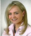
Dr. Dobrowolska has completed her PhD at the age of 27 years from Jagiellonian University Cracow Poland and University of Louisville USA. She is the Associate Research Scientist in Columbia University USA. Her research interests focuses on hematopoietic stem cells and novel markers in diagnosis and treatment of blood malignancies. The results of her work have been presented in numerous international meetings and publish in recognized journals.
Abstract:
Stem cells are the objects of scientific interest for many years. It is known that stem cells are the only cells in the body that can self-renew and differentiate to all type of the cells. Hematopoietic stem cells which can be received from bone marrow, peripheral blood or cord blood are used during auto- and allogenic transplantation in order for the reconstitution of bone marrow after mieloablative procedure. A lot of elements have influence on the way of hematopoietic cells preparation for transplantation, including conditions in which the cells are stored and prepared, knowledge and preferences of teams carrying out these activities. The protocols used for processing and storing stem/progenitor cells which are used as a transplant material are not unified around the world. Some factors like temperature, storage time and type of anticoagulants can have influence on preservations and cells’ survival and as a consequence have an impact on their ability for engraftment. As the number of hematopoietic cells transplantation increase constantly every year, the aim of my work is to optimize the hematopoietic cells storage conditions which help to maximize the hematopoietic stem cells ability for homing and reconstructing hematopoiesis after transplantation.
Wendy W. Weston
1VIVEX Biomedical
USA
Title: The RNA profile of exosomes from MSCs during osteogenic differentiation
Time : 15:35-15:55
Biography:
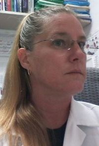
Wendy Weston is currently the Staff Development Scientist at VIVEX Biomedical. She completed her Postdoctoral studies at Bruce W Carter VA Medical Center, GRECC researching isolation of specific MSCs known as MIAMI cells. She completed her PhD from University of Miami, Miller School of Medicine in Molecular, Cell and Developmental Biology investigating the metastability of HSCs and the BM niche. She worked on bone formation and acceleration of fracture healing in the rat at Loma Linda VA and studied somatic gene therapy for muscle atrophy and B-MHC promoter activity following spinal cord isolation at Cal Poly, Pomona (MS Biological Sciences/Chemistry).
Abstract:
The importance of exosome contribution to the success of MSC healing properties has recently become clear. It has been suggested that exosomes are responsible for paracrine signaling to themselves and neighboring cells, resulting in immune mediation, tissue repair and differentiation into a variety of cell types, depending on the parental cell, time of collection and neighboring environment. We are interested in discovering the RNA components of the abundant exosomes produced by MSCs as they transition to osteoprogenitors. It is intuitive that the RNA excreted by the exosomes will differ at various stages of differentiation. There is already good evidence that the RNA composition of exosomes from cultures varies between cells and over time. Importantly, we wish to know if the changes in exosome RNA correlates with the signals necessary to drive the differentiation process and if previously undiscovered stimulants are present. If either previous or latter are found, it is possible that the purified exosomes from MSC culture supernatant at specific stages would elicit specific results upon transplantation. We are utilizing MIAMI cells exposed to osteogenic media for 21 days. Supernatant from the cell cultures are collected at day 0, 2, 7, 14 and 21. Exosomes are isolated from the supernatant and RNA is isolated from the exosomes. RNASeq is performed utilizing the Ion Proton Next Generation Sequencer. The RNA content of exosomes from each time point are being compared for elucidation of which remain constant and which change during various stages of osteogenic differentiation.
Christophe Thiriet
Université de Nantes
France
Title: The histone tail domains are critical in cell proliferation
Time : 16:10-16:30
Biography:
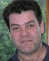
Christophe Thiriet has completed his PhD studies on chromatin in France and joined the Jeffrey J. Hayes Lab at the University of Rochester for his postdoctoral training on chromatin biochemistry. He has a permanent position at CNRS and he is the group head of the team ‘Epigenetics: proliferation and differentiation’ at the University of Nantes (France).
Abstract:
During cell proliferation, the genetic material and the epigenetic information need to propagate through daughter cells. A key step for these inheritances is the replication occurring in S-phase of the cell cycle. At this stage of the cell cycle, replication-coupled chromatin assembly of parental and newly synthesized histones is required for regaining the initial chromatin structure. Our group has developed a novel methodology to examine the function of the tail domains of histones in taking advantage of unique properties of the Physarum polycephalum model system. We showed that depending upon the histone complexes the tail domains did not present the same requirements. Indeed, while the tail domains of H2A/H2B are dispensable for nuclear import but required for chromatin assembly, the H3/H4 tail domains are needed for both nuclear import and chromatin assembly. We demonstrated that H3/H4 tail requirement is associated with histone acetylation. These results led us to propose a model showing the importance of well-conserved replication-dependent diacetylation of H4.
Itamar Barash
Agricultural Research Organization
Israel
Title: Delineating stem cells and cell hierarchy in the bovine mammary gland
Time : 16:30-16:50
Biography:
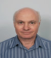
Itamar Barash has completed his Ph.D. at the age of 34 years from the Hebrew University of Jerusalem and Postdoctoral studies from McGill University, Polypeptide Hormone Laboratory. Montreal Canada, and the Weizmann Institute of Science, Molecular Genetics Department. Rehovot, Israel. He is Senior Research Scientist at the Volcani Center and a Lecturer in the Faculty of Agriculture and Environmental Sciences of the Hebrew University of Jerusalem. He has published more than 47 papers in reputed journals.
Abstract:
The bovine mammary gland is a favorable organ for studying mammary cell hierarchy due to its adaptation to extensive cell expansion, differentiation and renewal. The higher stem cell number at the onset of lactation may augment lactation persistency and add commercial benefits to the scientific knowledge. However, little is known about the bovine mammary stem cells and cell hierarchy. Here, bovine mammary epithelial cells were sorted according to the expression of CD24 and CD49finto 4 enriched populations: Stem cells, Basal cells, luminal progenitors and differentiated luminal cells. The enriched CD49fpos/CD24med population exhibited in vitro stem cell properties and held the greatest in vivo potential to regenerate the mammary epithelium, upon transplantation into the mammary fat pad. Clone formation, gene expression and floating mammosphere analysis combined with the transplantation experiments suggested that the stem cells give rise to bi-potent progenitors which differentiate along the basal/ myoepithelial lineage and possibly also give rise to luminal cells. The stem cells also generate luminal-restricted progenitors that give rise to differentiated luminal cells. Outgrowths generated by bovine cell transplantation maintained bovine cell characteristics, but did not expend in the mouse mammary fat pad. Fibroblast pre-transplantation improved outgrowth frequency, but not expansion. Limited expansion was achieved with estrogen and progesterone treatment. The underlying mechanism of the restricted bovine outgrowth development was studied in vitro. Apparently, it involves secreted factor(s) from the mouse mammary stroma that inhibit the development of foreign epithelial structure via paracrine signaling and may serve as a local defense mechanism against foreign tissue development.
Guido KRUPP
AmpTec GmbH
Germany
Title: Synthetic mRNAs as optimised tools for stem cell generation and for manipulating cellular phenotypes
Time : 16:50-17:10
Biography:
Dr. Guido Krupp (Ph.D.) is CEO and president of AmpTec GmbH. In 1981 he received his Ph.D. degree (Würzburg University & Max-Planck-Institute Martinsried). From 1983 to 1987 he was post-doc at Yale University. From 1987 to 2002: research group leader at Kiel University. Founder of artus GmbH (year 1998) & AmpTec GmbH (year 2005). Research interests: nucleic acid technology with focus on RNA, plant pathogens (viroids), ribozymes and telomerase. More than 60 publications, editor of Ribozyme Biochemistry & Biotechnology, and of Telomeres, Telomerases & Cancer, in the editorial board of Biotechnology Annual Review; SAB member of Orthogenics AS, Tromso, Norway.
Abstract:
Availability of high quality synthetic mRNAs (syn-mRNAs) has enabled progress in their applications. Important structural features, alternative technical options for high-amount, high-quality mRNA synthesis and GMP-compliant manufacturing and quality requirements are presented. Requirements in the application of mRNA-mediated manipulation of cells are presented (i) mRNA-directed expression of antigens in dendritic cells for vaccination projects in oncogenesis, infectious disease and allergy prevention; (ii) reprogramming of human fibroblasts to induced pluripotent stem cells with their subsequent differentiation to the desired cell type; (iii) applications in gene therapy.
Christopher A Bradley
Lattice Biologics, Inc., USA
Title: Extracellular matrix-assisted cell viability
Time : 17:10-17:30
Biography:
Christopher A. Bradley (Ph.D.), began his scientific research career in the field of Translational Control of Gene Expression at Louisiana State University Health Sciences Center in Shreveport, Louisiana. He has held several postdoctoral research positions within the University of California Davis Health System performing research in the fields of proteomics, head-and-neck cancer and osteoarthritis. His academic experience in Arizona involving virology and nucleocytoplasmic trafficking prepared him for industry positions in nutraceuticals and regenerative medicine. He currently serves in stem cell product development as Project Manager for Lattice Biologics, a certified tissue bank and manufacturer of surgical allografts.
Abstract:
Decellularized tissue matrices derived from cadaveric sources provide allografts used in surgical applications such as spinal fusion, tendon replacement, breast reconstruction, and dermal wound healing. The emergent technology of adult stem cells promises to augment these traditional surgical materials to create medical treatments that are truly regenerative. The next generation of allografts that will be self-healing and superior to previous implementations will require the application of stem cells and stem cell-derived technologies. In the case of bone tissue, osteoinductive properties of demineralized bone matrix are retained and can direct differentiation of recruited stem cells to promote healing. However, cell homing, retention, and vascularization at the lesion site could be greatly improved. Research to define the niche specified by stem cell-secreted extracellular matrix (ECM) has allowed for customization of the ideal healing microenvironment. Allografts infused with stem-cell derived ECM can potentially provide all the necessary signals that would improve engraftment and viability of recruited stem cells. Additionally, manipulation of the growth conditions under which stem cells produce their ECM could revive quiescent cells and enhance vascularization. Lattice Biologics seeks to develop the next generation of allografts. We envision using fluorescent chimeric RNA aptamers to measure important metabolite ratios in living cells and select for conditions ideal for autologous stem cell transplantation. Similarly, optimization of conditions used to grow stem cells in vitro could influence the output of soluble and insoluble signals present in secreted ECM which would then be used to instruct a patient’s own circulating stem cells.
Shaohua Li
Rutgers-The State University of New Jersey
USA
Title: CREG1 interacts with Sec8 to promote cardiomyocyte differentiation of embryonic stem cells
Time : 17:30-17:50
Biography:
Abstract:
CREG1 is a small glycoprotein originally identified in a yeast two-hybrid screen of the Drosophila cDNA library. To investigate its physiological functions, we first performed immunostaining and immunoblotting on mouse tissues. We found that CREG1 is highly expressed in both embryonic and adult hearts. During mouse embryonic stem (ES) cell differentiation into cardiomyocytes, the expression level of CREG1 increases and correlates with myogenic differentiation. Overexpression of CREG1 in ES cells enhances cardiomyogenesis, whereas knockout of CREG1 has an inhibitory effect. Chimeric analysis suggests that the cardiac-inducing activity of CREG1 is cardiomyocyte intrinsic. Affinity pull-down assay using His-tagged CREG1 followed by mass spectrometry identified a novel interaction between CREG1 and Sec8 of the exocyst complex essential for tethering vesicles to the plasma membrane. In vitro protein binding assay using recombinant His-tagged CREG1 and the Sec8-GST fusion protein confirmed direct binding of CREG1 to Sec8. Furthermore, site-directed mutagenesis revealed that the amino acid residues D141 and P142 in CREG1 are required for Sec8 binding. Rescue of CREG1 knockout ES cells with human wild-type CREG1 restores cardiomyocyte differentiation while the Sec8-binding mutant fails to do so. In addition, shRNA-mediated knockdown of Sec8 in wild-type ES cells also inhibits cardiomyogenesis. These results suggest that CREG1 binding to Sec8 promotes cardiomyogenic differentiation. Mechanistically, CREG1, Sec8 and N-cadherin are co-localized to both intercellular junctions and Rab11-positive recycling endosomes. Overexpression of CREG1 increases the N-cadherin protein level and enhances the assembly of adherens and gap junctions. By contrast, knockdown of Sec8 leads to cadherin degradation. Taken together, these data support a notion that CREG1 binds to Sec8 and enhances the assembly of intercellular junctions, thereby promoting cardiomyogenesis.
Prwati Armand
Airlangga University
Indonesia
Title: The Potential Differentiation of Hypoxic Niche Bone Marrow Derived Autologus Mesenchymal Stem Cells For Retinal Disorder Treatment
Time : 17:50-18:10

Biography:
Prwati Armand is a scientist, researcher currently working at Fakultas Kedokteran, Universitas Airlangga. She has experience in the areas of stem cell , regenerative medicine, bone marrow transplantation, pancreatic stem cells and diabetic treatment. At now, she has published more than 10 papers in reputed journals.
Abstract:
The use of stem cells in the treatment of retinal degenerative disease has received increasing attention recently . Based on embryogenesis retina cell was developed from neural crest, this is part of ectoderm layer. The neuroretina is a complex structure whose health depends on blood vessels and retinal pigment epithelium (RPE). Replacement therapy is an approach to regenerative medicine in which healthy cells replace cells that have died or are dysfunctional. For example, in retinitis pigmentosa, photoreceptors die. Replacement therapy for RP could involve transplantation of cells that can integrate with the host retina and function as photoreceptors. Replacement retinal therapy is sight-restoring also in diabetic retinopathy. To be useful for cell replacement therapy, stem cells must proliferate extensively to generate sufficient quantities of material if they are intended to serve as a “universal donor.” If the stem cells are derived from the patient (e.g., iPSCs), then the requirement for extensive proliferation may be reduced considerably since these cells will serve only one recipient. Stem cells must stably differentiate into the desired cell type(s). ESCs and iPSCs vary in their tendency to differentiate into cells of a given lineage . MSCs are multipotent, which means they can form multiple cell types of one lineage. For example, a retinal progenitor cell can give rise to photoreceptors, bipolar cells and ganglion cells but not to corneal cells. Adult stem cells may remain quiescent for long periods until activated by a normal need for more cells to maintain tissues . To extend the potential tendency differentiated of bone marrow derived autologus MSCs to the retinal cells we make a quiescent condition of the MSCs by niche modification of culture with hypoxia before to injection to the patients. We measure the express of Octamer 3/4 (Oct4), sex determining region Y box–containing gene 2 (Sox2), and Nanog as a marker of pluripotency and also evaluate the optimal passage of stem cell before used.
Reinaldo Geraldo
Federal University of Juiz de Fora, Brazil
Title: Platelet Activity: The assessment profi le in patients with sicle-cell hemoglobinopathy and the antiplatelet potential of synthetic compounds
Time : 18:10-18:30

Biography:
Reinaldo Barros Geraldo is a young scientist, researcher currently postdoc at Federal University of Juiz de Fora (Brazil). Graduated in Biomedicine Fluminense Federal University (2007), Master of Neuroimmunology, Fluminense Federal University (2009) and Doctor of Science by Federal University of Rio de Janeiro (2013). Has experience in the areas of biochemistry, with an emphasis on computer modeling, acting on the following subjects: Comparative modeling, structural biochemistry and Molecular Biology. At now, he has published 11 papers in reputed journals.
Abstract:
Platelets and erythrocytes are blood elements involved in important pathologies such as the hemoglobinopathies, thrombosis and occlusive processes, and they may be both correlated to them. The treatment of these pathologies is a science purpose as the current options present risks and collateral effects. This work purpose was to produce a review about platelets and erythrocytes and their participation in some pathology, also describing a brief study about these elements and a hemoglobinopathy patient. We also identified some molecules with an antiplatelet profile on rabbit blood from three new acylhidrazones and pirazole-piridine serie synthetized by Alice Bernardino and Vitor Ferreira groups from Instituto de Química da UFF (Labiomol AM serie) and Luiza Dias e Maria Abadia Di Vaio groups from Faculdade de Farmácia da UFF (Labiomol LHa e Labiomol LHb serie), using ADP, arachidonic acid and collagen as agonists. The LabiomolLHa serie was the most promising on platelet assay induced by arachidonic acid, where Labiomol LHa 41, 43, 62, 65, 66, 67 e 130 compounds were able to totally inhibit the platelet aggregation.

