Day 3 :
- Workshop on Synthesis of scaffold for heart valve based on Molecular patterns and study of its effects (Mechanically، Biologically، Collagen 1 and Elastin Genes Expression) on Valvular Interstitial Cells
Location: Vienna Room
Chair
Maryam Eslami
Harvard Medical School, USA
Session Introduction
Maryam Eslami
Harvard Medical School
USA
Title: Synthesis of scaffold for heart valve based on Molecular patterns and study of its effects (Mechanically، Biologically، Collagen 1 and Elastin Genes Expression) on Valvular Interstitial Cells
Time : 08:55-09:20
Biography:
Eslami graduated with MD (2010), PhD (2014) degrees. She has joined in Harvard Medical School (Harvard-MIT Division of Health Sciences and Technology) as a part of her Ph.D. dissertation in the field of Heart valve. She has carried out some broad research on Orthopedic fractures and has published a book and papers in this field. Her U.S and PCT patent achieved the rank of “Best 2008 Invention†from WIPO (World Intellectual Property Organization of the United Nations) and she has received the title of "Best 2008 Women inventor" from WIPO and 6 Gold Medals and 6 Honorary Diplomas in the Contests and Fair of the Inventors in Geneva and South Korea. International and national awards for her research were one of the fundamental achievements that she has received. Now she is a research vice president of Tehran Paramedical School, Azad University.
Abstract:
The incidence and prevalence of heart valve disease is increasing worldwide and the number of heart valve replacements is expected to increase significantly in the future. By mimicking the main tissue structures and properties of human heart value, tissue engineering offers new options for the replacements. Applying an appropriate scaffold in fabricating tissue engineered heart valves (TEHVs) is of importance since it affects the secretion of the main extracellular matrix (ECM) components, collagen 1 and elastin, which are crucial for providing the mechanical, elastic and tensile strength of TEHVs.Using biliogical, mechanical and Real Time PCR experiments, the relative collagen 1 and elastin genes expression levels obtained for three samples of each examined valvular interstitial cells (VICs)-seeded scaffolds including electrospun poly(glycerol sebacate) (PGS)-poly (ε-caprolactone) (PCL) microfibrous scaffolds, methacrylated gelatin (GelMA) and methacrylated hyaluronic acid (HAMA) based hydrogel-only and the composite (consists of PGS-PCL and hydrogel) scaffolds. Sheep mitral valvular interstitial cells were encapsulated in the hydrogel and evaluated in hydrogel-only, PGS–PCL scaffold-only, and composite scaffold conditions. Although the cellular viability and metabolic activity were similar among all scaffold types, the presence of the hydrogel improved the three-dimensional distribution of mitral valvular interstitial cells. As seen by similar values in both the Young’s modulus and the ultimate tensile strength between the PGS–PCL scaffolds and the composites, microfibrous scaffolds preserved their mechanical properties in the presence of the hydrogels. Our results showed that the level of relative expression of collagen and elastin genes was higher in the VICs- encapsulated composite scaffolds compared to PGS-PCL-only and hydrogel-only scaffolds and the difference was statistically significant (P < 0.05). The maximum difference of elastin and collagen 1 genes expression was between the composite scaffold and the hydrogel-only scaffold, with the most and the least quantity, respectively.The VICs-encapsulated composite scaffold was observed to be more inductive to ECM secretion over the PGS-PCL-only and hydrogel-only scaffolds. This composite scaffold can serve as a three-dimensional structure model for heart valve tissue engineering with the capability of providing the necessary mechanical-properties such as, elasticity and tensile strength and the ability to grow, repair and be remodelled as a tissue.
- Track 2: Drugs and clinical Developments
Track 14: Signalling in Development
Track 17: Genetics, Bioinformatics and Computational Biology
Location: Vienna Room
Chair
Diana Anderson
University of Bradford, United Kingdom
Session Introduction
Kazuo Ishii
Tokyo University of Agriculture and Technology
Japan
Title: Statistical computing and mathematical modeling for understanding of biological functions using big data
Time : 09:20-09:40
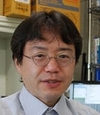
Biography:
Kazuo Ishii, Ph.D. is the Professor of Genomic Sciences, the Graduate School of Agriculture, Tokyo University of Agriculture and Technology. After receiving the PhD degree from the University of Tokushima Graduate School of Medicine in 1995, he began the professional career in genomic sciences. He has studied data analysis and data mining for microarray and next generation sequencing since 2003. His present interest is statistical computing and mathematical modeling for biology
Abstract:
Emergence of the next generation sequencing technologies and its application to clinical and biological research opened the door of the era of genomic big data. Today, realizing the personal medicine with big data is fascinating topic in the current clinical research. For manipulation and processing of genomic big data, we are utilizing four strategies of information technology: (1) Monte-Carlo Simulation, random sampling from the genomic big data. (2) HPC (high performance computing), possessing Many Core CPU and Large Memory. (3) Cloud-based Hadoop technology containing distributed file systems (HDFS) and distributed processing systems (MapReduce) on the Amazon Web Services (AWS). (4) non-Hadoop Shell scripting technology including distributed file systems and distributed processing system without hadoop. Monte-Carlo Simulation is a powerful method for optimization and elucidation of the biological functions using genomic big data. Here we will focus on the powerful and novel application of Monte-Carlo Simulation for genomic big data.
Julie R. Murrell
EMD Millipore, Bedford, MA
USA
Title: UPSTREAM EXPANSION SOLUTIONS FOR STEM CELLS
Time : 09:40-10:00
Biography:
Dr. Julie Murrell is a Senior R&D Manager for Stem Cell Biology and Collaborations at EMD Millipore. Dr. Murrell has led an early technology assessment group for the past 7 years and has been part of the Stem Cell group for 3 years. Through that time, she has led the efforts to establish robust assays and identify new targets as key quality attributes for large scale stem cell manufacturing, with a special focus on hMSCs. Dr. Murrell’s background is in Cell and Molecular Biology. Her multi-disciplinary background has led to innovative team-driven approaches in the field of stem cell production.
Abstract:
The long-term view of regenerative medicine therapies predicts an increased need for expansion solutions that ease scalability, utilize animal origin-free materials and are compatible with limited downstream processing steps. As more stem cell therapeutics progress through clinical testing, current in vitro culture methods in 2D vessels are proving cumbersome to scale. Moreover, the concurrent decreased demands for serum from the recombinant protein and vaccines markets may result in a shortage of serum as clinical cell therapy programs are successful. Currently available technologies and challenges in the upstream bioprocessing space, focusing on expansion of allogeneic mesenchymal stromal/stem cells (MSCs) will be reviewed within the context of specific case studies. First, we have developed an approach for selecting media and microcarriers, using adipose-derived MSCs as a model cell line. Next, an evaluation of animal-free media supplementation and cellular detachment solutions was performed. Because cellular therapeutic manufacturing processes are further complicated by the requirement to separate cells from microcarriers whilst retaining cell yield, viability and target phenotypic and functional characteristics, the importance of considering downstream challenges while establishing upstream parameters will be highlighted.
Stephen Lin
Thermo Fisher Scientific
USA
Title: Proximity ligation assays, A method for ultra sensitive protein detection
Time : 10:00-10:20

Biography:
Stephen Lin received his Ph.D. from Washington University in St. Louis in 2002 and did his postdoctoral research at Harvard University. In 2006 he joined StemCells,Inc of California as a scientist for liver cellular therapeutics, where he discovered pathways that contribute to the rapid decline of function and expansion of primary human hepatocytes. Since 2012 he has been staff scientist for early-stage product concepts at Life Technologies (now part of Thermo Fisher Scientific), a global life sciences company that develops and offers tools for every aspect of stem cell and gene therapy including genetic manipulation, genetic analysis, and cell culture.
Abstract:
Quantitative PCR (qPCR) has revolutionized the study of cellular nucleic acids, including genomic DNA, mRNA and micro RNAs. Now TaqMan® Protein Assays, which are based on the proximity ligation assay (PLATM) technology, extend qPCR applications to the analysis of proteins through the amplification of a surrogate DNA template. This technology has been optimized for use with crude cell lysates, tissue homogenates, and blood samples and has been combined with TaqMan® chemistry to create a highly sensitive and specific process for measuring proteins. With its simple and rapid workflow akin to traditional qPCR techniques, PLA enables a broad range of applications that includes the analysis of proteins from complex or difficult samples such as serum and plasma, protein modifications such as phosphorylation, and protein and gene expression from a single sample and even a single cell in parallel on the same platform. Moreover, PLA is being applied in new ways to study normal and aberrant biological processes that are associated with homeostasis, growth and differentiation. The addition of TaqMan® Protein Assays to the qPCR assay repertoire marks the beginning of a new era in the analysis of proteins that will ultimately provide a better understanding of biological systems.
Michal K. Stachowiak
State University of New York, USA
Title: Evidence based theory for an integrated genome regulation in ontogeny and its potential therapeutic targeting
Time : 10:20-10:40

Biography:
Michal K. Stachowiak has completed his Ph.D from the Gdansk Medical University, Poland, in 1980, and postdoctoral studies at the University of Pittsburgh. He is director of the Molecular and Structural Neurobiology and Gene Therapy Program and Western New York Stem Cell Engraftment and In Vivo Analysis Core. He has published more than 100 papers in reputed journals. His book: “Stem Cells - from Mechanisms to Technologies” (World Scientific Publishing) describes fundamanetal mechanisms in the biology of stem cells and their terapeutic utilization.
Abstract:
The advent of multicellular mechanisms required an imposition of new controls over the natural propensity of single cell organisms to proliferate, invade new niches and transmit their genetic material. A strict dominion over the cell proliferation, migration and differentiation had been imposed so that the complex tissues and organs may be formed. The construction of the metazoan body engages myriads of genes and gene–controlling transcription factors (TFs). Their actions need to be coordinated in the context of 3D chromatin structure to ensure programmatic realization of the genetic blueprint. How such an immense task is accomplished remains largely unknown. Our studies have revealed a pan-ontogenic gene regulation mechanism, Integrative Nuclear FGFR1 Signaling (INFS), which controls pluripotency, cell cycle and differentiation programs. At its center are proteins that bear the historic name of Fibroblast Growth Factors (FGF) and FGF receptors (FGFR). Neither FGFs nor FGFRs exist in single-cell organisms, but are common to eumetazoans and key for the generation of specialized tissues. Gene ablation experiments demonstrated that mammalian fgfr1 and its invertebrate orthologs are essential for early gastrulation, development of mesodermal somites, nervous system (etc.), by affecting “master” genes of various ontogenic networks including Notch, Wnt, Hox, SOX, (etc.), and microRNAs. FGFs have emerged during early metazoan evolution equipped with nuclear localization signals (NLS), acting as nuclear proteins. Various members of the mammalian FGFs family retained NLS, likewise, individual FGFR (i.e. FGFR1) have structural adaptations that allow an accumulation and action directly in the cell nucleus. Most recently, RNAseq and ChIPseq have delineated a global and direct gene programing by nFGFR1, ensuring that pluripotent embryonic stem cells (ESCs) develop into multipotent neural progenitor cells (NPCs) and their further differentiation. nFGFR1 alone, and with its partner RXR and orphan receptors, targets promoters of thousands of genes, and forms multi-gene chromatin topological domains and controls the expression of pluripotency, cell cycle, ectodermal, morphogenic, neuronal, and mesodermal genes [6]. nFGFR1 targets genes in major developmental pathways and consensus sequences of TFs encoded by nFGFR1 targeted genes. nFGFR1 targets miRNA genes as well as DNA insulator sequences and is involved in active topological chromatin domains (multi-gene transcription hubs). These findings define previously unknown global and hierarchical genome control, which enables ontogeny and offers a new perspective on developmental disorders, cancer and their treatments. Such strategies may involve gene transfer-based and pharmacological re-activation of INFS to restore developmental-like neurogenesis in the adult brain.
Patricia Berg
George Washington University Medical Center
USA
Title: BP1, a Potential Oncogene Overexpressed in Cancer
Time : 10:55-11:15
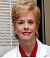
Biography:
Abstract:
BP1, a gene we identified and cloned, is a member of the homeobox gene family of transcription factors (TF). BP1 is overexpressed in breast cancer, prostate cancer, ovarian cancer, acute myeloid leukemia, non-small cell lung cancer, and possibly other malignancies as well. Important characteristics of BP1 in breast cancer include: (1) BP1 is expressed in 80% of invasive ductal breast tumors, including 89% of the tumors of African American women compared with 57% of the tumors of Caucasian women. (2) BP1 expression correlates with the progression of breast tumors, from 0% in normal breast tissue to 21% in hyperplasia and 46% in ductal carcinoma in situ. (3) Expression of BP1 is associated with larger tumor size. (4) BP1 appears to be associated with metastasis. Forty-six cases of inflammatory breast cancer were examined and all were positive for BP1 expression, as well as matched lymph nodes in the nine metastatic cases. (5) BP1 overexpression induces oncogene expression, including BCL-2, VEGF and c-MYC, as well as other genes important in angiogenesis, invasion and metastasis. pBP1 down-regulates BRCA1. (6) BP1 up-regulates ER alpha and induces estrogen independence. High pBP1 levels can lead to estrogen independence in ER positive breast cancer cells and tumors in mice. In summary, BP1 appears to confer properties on breast cancer cells that lead to a more invasive and aggressive phenotype. Since the functions of homeotic TF are highly conserved, it is likely that BP1 regulates many of the same processes and genes in other malignancies.
Manuela Stoicescu
Oradea University
Romania
Title: Beta-Human Chorionic Gonadotrophin (β-HCG total) as a tumor marker in pregnancy
Time : 11:15-11:35

Biography:
Manuela Stoicescu, Consultant Internal Medicine Doctor (PhD in Internal Medicine) now is Assistant Professor of Medical Disciplines Department, University of Oradea, Faculty of Medicine and Pharmacy, Romania, Internal Medicine Hospital and Office. She is Member of Romanian Society of Internal Medicine, Member of Romanian Society of Cardiology, Chemistry, Biochemistry and Member of Balcanic Society of Medicine. She was invited as a speaker at 23 International Conferences, she is Editorial Board Member in three ISSN prestigious Journal in U.S.A, she published 12 articles in prestigious ISSN Journals in U.S.A. she published four books: two books for students in English and Romanian language:” Clinical cases for students of the Faculty of Medicine”, one book in English language on Amazon at International Editor – Lambert Publishing Academic House in Germany- “Side Effects of Antiviral Hepatitis Treatment”, one monograph in Romanian language” High blood pressure in the young a ignored problem!” two chapter books – Cardiovascular disease: Causes, Risks, Management CVD1- Causes of Cardiovascular Disease 1.5,1.6, U.S.A on Amazon.
Abstract:
Beta-Human Chorionic Gonadotrophin (β HCG total) represents a substance that is usually secreted by the placenta in the course of pregnancy. This is why it is used as a pregnancy test, because the values are high in the first trimester of the pregnancy. But the determination of the β HCG total is also can used as a tumor marker in order to detect gestational trophoblast diseases such as the hydatidiform mole and choriocarcinoma and germ cell tumors of the ovaries and testicle. Nevertheless the young pregnant hypertensive patient, with increased values of total β HCG after the first trimester of pregnancy, cannot be left to be interpreted only within the pregnancy context, but these must be taken into account as a possible choriocarcinoma diagnosis. This tumoral marker should be performed as a screening test in young pregnant women with severe values of blood pressure, after the first month of pregnancy, in order to detect an unknown choriocarcinoma, thus the blood pressure values should not be interpreted only within the pregnancy context, of course if other causes of secondary hypertension in pregnant woman were excluded. In the future the stem cell therapy it is posible to be a solution for this patients with choriocarcinoma unsolved yet.
Abdulkader Rahmo
New Cell Sciences, Inc
USA
Title: Relevance and scope of SMS derived Multicellular Micro Machines (M3) in Native tissue reconstruction.
Time : 11:35-11:55
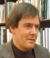
Biography:
Abdulkader Rahmo has completed his Diploma in Biochemistry in 1988 at the Swiss Federal Institute of Technology (ETHZ), his Ph.D in Biochemistry and Molecular biology in 1994 at the University of Southern California (USC) in los Angeles USA. He was heading the medical biotech section at the National Commission for Biotechnology. Currently he is the CSO of a newly established American biotech company. He is sole author of several scientific books and coauthor of many scientific articles.
Abstract:
The newly discovered Small Mobile Stem (SMS) cells exhibit the extraordinary ability to form diverse complex cooperative multicellular assemblies. These assemblies have a demonstrated aptitude for engineering tissue like structures in an in vitro cell culture set up. The observed approach to construction can be ascribed to a top down modus operandi at a micro level. It is executed by units we designated as Multicellular Micro Machines (M3). To explore the relevance and scope of this in vitro discovery, for the in vivo case of tissue reconstruction, M3 assemblies and their traces were hunted in a broad variety of native tissues. Results indicate the importance of implicating the M3 mediated mechanism in native tissue reconstruction; and demonstrate hence the fundamental role of SMS cells.
Akshay Anand
Post Graduate Institute of Medical Education and Research India
Title: Does hUCB derived Lin-ve stem cells rescue amyloid-β peptide induced mouse model of memory loss
Time : 12:15-12:35

Biography:
Akshay Anand graduated from Post Graduate Institute of Medical Education and Research (PGIMER). He is interested in understanding the molecular mechanisms of neurodegeneration utilizing invitro, invivo, alternative and biotherapeutic approaches. He has been involved in the discovery of animal model of AMD published in Nature Medicine. Dr Anand is a recipient of ICMR Shankuntla Amir Chand Award (2010), Young Scientist Award from DAE (2005), Runners up for NASI Scopus Award for Biological Sciences(2012), served as Judge at ISEF, USA (2002-3), and is the Editor in Chief of Annals of Neurosciences.
Abstract:
Objectives: The objective of the study was to evaluate the effect of lineage negative human umbilical cord blood derived stem cells in rescue of memory loss in animal model of AD.
Methods: We used digital stereotaxis, immunohistochemistry, real time PCR and behavioral assays such rota rod and morris water maze in order to evaluate the learning and memory in experimetal and control Swiss Albino mice. Lineage negative stem cells were purified by MACS and labelled by CFDA dye in order to trace the stem cell recruitment, incorporation and differentiation and FACS analysis to characterise these cells before transplantation.
Results: There was dose dependent rescue of memory loss caused by transplantation of lineage negative hUCB cells. There was increase in BDNF and CREB expression subsequent to transplantation of Lineage negative human UCB cells as well as decline in apoptosis analysed by Fas L and Fluro Z. The immunolocalisation of Abeta reduced with transplantation of these cells.
Conclusion: Lineage negative UCB cells possess the necessary neurotrophic effect in extending the survival of hippocampal neurons which may contribute in rescue of memory loss. The immunolocalisation of Abeta reduced with transplantation.
Leigh J. Mack
CIMTESES Foundation, USA
Title: New Intercellular Surgical Closure System
Time : 12:35-12:55

Biography:
Leigh J Mack completed his MD and PhD (March, 2015) at USAT Montserrat with a post-graduate training at University of Oxford in Nanotechnology for Medicine. He is the President of CIMTESES Foundation, Curacao. He is also is the Chief Medical Officer for Inaline, Corp. In addition, he is a consultant for several other companies and on the Board of Directors for many start up companies.
Abstract:
A new approach to surgical closures using a three-dimensional, intercellular space attachment system to approximate tissue. The design is mechanical in nature and does not employ adhesives. The system interfaces directly will the cellular wall to form attachments at the micron and nano level. The system also employs multiple modalities of attachment to provide hundreds of secure points of attachments for a highly effective hold.
Nasrin Moazami
Iranian Research Organization for Science and Technology
Title: A novel metabolite from an extremophile Streptomyces with apoptosis regulator function
Time : 12:55-13:15
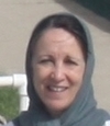
Biography:
Abstract:
Kavir-48, a protein with a molecular weight of 44 KDa., isolated from an extremophile Streptomyces was investigated for anti proliferative activity on 10 human cell lines. 5000 cells were plated in 96 well plates. After 24 h. Kavir -48 was added in five dose levels (0.01-100 µg/ml). After 4 days of continuous exposure, the cellular DNA content and morphology was determined, using propidium iodide staining, the resulting fluorescence correlates with a number of leased cells. A selective growth inhibition (Mean IC 70) observed for the large cell Lung cancer cell line and mammary cell line. The antitumor activity as well as tolerability of product was evaluated in In-vivo testing on lung cancer cells, (Xenograft in nude mice) since there might be a selective antitumor effect after 21 days of administration .Neither lethality nor body weight loss was observed in all group of tested animal, and in the large cell lung cancer model, a clear anti tumor effect observed after only five days of administration. Amino acid sequencing of protein suggest that the compound consist of a fusion proteins NISOD and a Tat protein combination. Expression of the enzymatic activity of the transduced Tat-SOD fusion proteins is essential for therapeutic application; for several reasons, such as the size and instability of the SOD enzyme these attempts to develop the natural enzyme for clinical use have been largely unsuccessful.
Award Ceremony
Closing Ceremony
Shivdas G Nanaware
D. Y. Patil University
India
Title: FORMED CELLULAR ELEMENTS OF BLOOD-PLATELETS: EXCELLENT WOUND HEALING CELLULAR TREATMENT

Biography:
Prof.Dr.Shivdas Ganpati Nanaware is a Dean, Faculty of Interdisciplinary studies and Co-ordinator, Stem Cell and Regenerative Medicine Department at Center for Interdisciplinary Research, D.Y.Patil Unviersity, Kolhapur, Maharashtra,India. Prof. Nanaware completed his Ph.D. at the age of 30 years from Shivaji University Kolhapur.After completion of his Doctorate he has served in the same University as a Professor and Head, Department of the Zoology for 35 years. Prof.Nanaware has a unique combination of rich scientific research and administration experience. His research is related to cellular changes in response to water polluting toxicants, pesticides and plant derived bioactive compounds. His research work on crop distructive molluscan pest snails and slugs is well recognized by the researchers working in Malacology.He has developed a “Confirmatory Metachromatic Histochemical Technique for the detection of unusual Polysaccharide, Galactogen†in animals. He has published more than 60 papers in reputed generals and has guided 20 research scholars for their M.Phil. and Ph.D. degrees. He is a member of editorial boards of many Journals of cell and tissue research. He has attended several national and international conferences in India and abroad. Prof.Nanaware is a life member of many professional societies. He has received several awards and honors in recognition of his meritorious research work. He is a recipient of Best Teacher Award, Govt. of Maharashtra and Shivaji University, Kolhapur. He is also a Gold Medal Awardee of Academy of Environmental Biology. Dr.Nanaware has interest in many branches of Zoology, Toxicology, Neurobiology, Cell biology Stem Cell and Regenerative Medicine and Clinical Application of Cell Therapy.
Abstract:
The wound management forms an important part of the medical and pharmaceutical concern worldwide. Many methods were traditionally used to cure wounds including dressing with natural or synthetic bandages such as cotton, wool, lint and gauzes, and the extracts of plant parts, oils and ghee. The effective wound management depends on understanding a number of different factors such as the type of wound, the healing process , condition of the wound in terms of health and infection ( e.g. diabetic) environment and social setting and the physical and chemical properties of the available dressings. In recent advances it has been proved that the platelets, one of the blood cellular elements, play important role in the wound healing mechanism both in wounds and non-healing diabetic wounds. Platelet cells are practically involved in every event of the wound healing cascade including homeostasis, proliferation, remodeling and cell signaling. Platelet cell growth factors like PGDF, TGF Alfa , TGF Beta and EGF help in chemotaxis, fibroblast proliferation, collagen production and metabolism, epithelial cell proliferation and granulation and tissue formation related functions in wound healing. With the development of platelet cell based procedures at our center such as use of activated platelets, platelet riched plasma (PRP), polymer based scaffolds of Polyvinyl alcohol (PVA) and Polyethylene glycol (PEG) and their combinations have showed dramatic and interesting changes during wound healing. Our efforts in developing the human skin engineering, cellular matrices and cell based therapies are in progress to improve perfect wound healing process. We hope that the potential application of these technologies will be helpful to the people with wounds in general and non-healing diabetic wounds in particular in the near future and will insure rapid and complete healing key strategy for the wound management.
- Genetics, Bioinformatics and Computational Biology


